
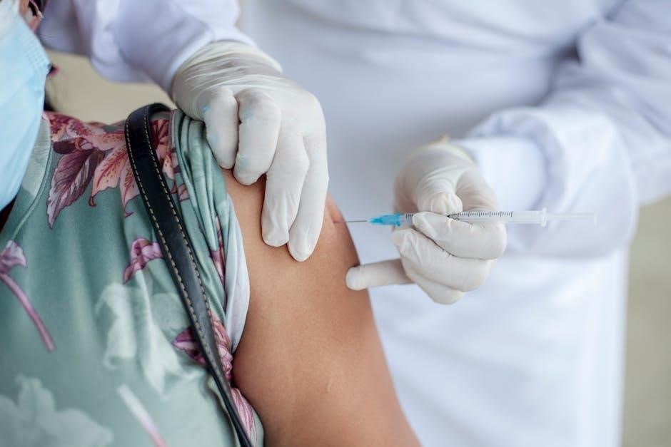
Fluoroscopy-guided injection is a medical imaging technique using real-time X-ray visualization to guide precise needle placement for administering medications or anesthetics. It enhances accuracy in pain management, spinal procedures, and joint injections, minimizing complications.
1.1 Definition of Fluoroscopy-Guided Injection
Fluoroscopy-guided injection refers to a minimally invasive medical procedure that utilizes real-time X-ray imaging to accurately guide the placement of needles or catheters during the administration of therapeutic agents, such as corticosteroids, anesthetics, or pain-relieving medications. This technique allows clinicians to visualize internal structures dynamically, ensuring precise delivery of the injectable material to the targeted tissue or joint. It is commonly employed in pain management, spinal interventions, and soft tissue injections, where anatomical accuracy is critical to achieving optimal therapeutic outcomes. The use of fluoroscopy enhances the safety and efficacy of the procedure by reducing the risk of misplacement and complications. Additionally, contrast agents are often used to improve visualization, enabling clearer delineation of the target area. This method is particularly valued for its ability to provide immediate feedback, making it a cornerstone in image-guided interventions.
1.2 Historical Development of Fluoroscopy-Guided Techniques
Fluoroscopy-guided injection techniques have evolved significantly since the discovery of X-rays by Wilhelm Conrad Röntgen in 1895. Early applications of fluoroscopy in the 20th century focused on diagnostic imaging, with limited use in interventional procedures. The 1950s and 1960s saw the introduction of image intensifiers, improving visualization and paving the way for guided injections. By the 1980s, fluoroscopy became a cornerstone in pain management, particularly for spinal and joint interventions. Advances in technology, such as digital fluoroscopy and better contrast agents, enhanced precision and safety. Modern systems now incorporate real-time imaging, reducing radiation exposure and improving outcomes. The historical progression underscores a commitment to refining techniques for accurate and minimally invasive treatments, laying the foundation for contemporary applications in various medical specialties.
Clinical Applications of Fluoroscopy-Guided Injections
Fluoroscopy-guided injections are widely used in pain management, spinal procedures, joint injections, and soft tissue interventions. They enhance precision, reduce complications, and improve outcomes in treating chronic pain and inflammatory conditions.
2.1 Pain Management and Fluoroscopy-Guided Injections
Fluoroscopy-guided injections play a pivotal role in pain management, particularly for patients with chronic pain conditions. By enabling real-time visualization, fluoroscopy allows precise placement of needles for administering anesthetics, corticosteroids, or other therapeutic agents directly to the pain source. This technique is commonly used in procedures such as epidural steroid injections, facet joint injections, and nerve blocks, where accuracy is critical to ensure effectiveness and minimize risks. The use of fluoroscopy in pain management reduces complications associated with blind or unguided injections, such as nerve damage or medication misplacement. It is especially beneficial for targeting complex anatomical structures, such as spinal facets or nerve roots, where precision is paramount. Patients often experience significant pain relief, improved mobility, and enhanced quality of life following these procedures. Fluoroscopy-guided injections are considered a gold standard in interventional pain management due to their high success rates and safety profile.
2.2 Role in Spinal Procedures
Fluoroscopy-guided injections are integral to various spinal procedures, offering precise visualization for targeting specific anatomical structures. This technique is widely used in epidural steroid injections, facet joint injections, and nerve root blocks, ensuring accurate needle placement; Real-time imaging allows practitioners to navigate the complex spinal anatomy safely, minimizing risks of nerve damage or incorrect medication administration. Fluoroscopy is particularly valuable in procedures involving the cervical, thoracic, and lumbar regions, where misplacement could lead to serious complications. Its use enhances the efficacy of pain relief interventions, such as in cases of herniated discs, spinal stenosis, or spondylolisthesis. By providing clear, dynamic images, fluoroscopy-guided injections enable physicians to deliver therapeutic agents directly to the source of pain, improving outcomes and reducing recovery times for patients with chronic spinal conditions.
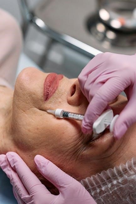
2.3 Use in Joint and Soft Tissue Injections
Fluoroscopy-guided injections are widely utilized in joint and soft tissue procedures to ensure precise needle placement and optimal therapeutic delivery. This technique is particularly beneficial for injections in large joints, such as the shoulder, hip, and knee, where accurate targeting is critical. Fluoroscopy allows real-time visualization, enabling practitioners to administer corticosteroids, anesthetics, or hyaluronic acid directly into the joint space or surrounding soft tissues. It is also invaluable for injections in smaller joints, like those in the hands or feet, where anatomy is complex. Additionally, fluoroscopy guides injections for conditions such as tendinitis, bursitis, and ligament sprains, ensuring medication reaches the intended site. The use of contrast agents further enhances accuracy, confirming proper placement before administering the therapeutic agent. This minimally invasive approach reduces the risk of complications and improves outcomes for patients with joint-related pain or inflammation, making it a cornerstone in modern orthopedic and rheumatologic care.
Equipment and Technology Used in Fluoroscopy-Guided Injections
Fluoroscopy-guided injections rely on advanced imaging equipment, including fluoroscopy machines, image intensifiers, and contrast agents. Modern systems incorporate radiation shielding, high-resolution monitors, and digital recording capabilities to enhance precision and minimize radiation exposure during procedures.
3.1 Fluoroscopy Machines and Their Features
Fluoroscopy machines are central to fluoroscopy-guided injections, utilizing real-time X-ray imaging to visualize internal structures. Modern systems often feature C-arm designs, allowing for flexible positioning around the patient. High-resolution image intensifiers enhance clarity, while digital imaging systems enable precise recording and playback. Many machines now incorporate advanced dose monitoring to optimize radiation exposure. Additional features include adjustable frame rates for varying procedural needs and integrated radiation shielding to protect both patients and operators. Some systems also offer advanced imaging modes, such as pulsed fluoroscopy, to reduce radiation further while maintaining image quality. These machines are equipped with high-resolution monitors, providing clear visualization for accurate needle placement. Together, these features ensure efficient, safe, and precise guidance during injections, making fluoroscopy machines indispensable in modern interventional procedures.
3.2 Contrast Agents in Fluoroscopy-Guided Procedures
Contrast agents play a critical role in fluoroscopy-guided injections by enhancing the visibility of anatomical structures. These agents, typically iodine-based or gadolinium-based, are administered to delineate soft tissues, joints, or spinal spaces. In procedures like epidural steroid injections or joint aspirations, contrast agents help confirm needle placement and ensure medication delivery to the target area. Iodinated contrast is commonly used for its ability to block X-rays, providing clear images of injected regions. Gadolinium-based agents are sometimes employed in combination with MRI for complex cases. Safety is paramount, as some patients may have allergies or renal impairment, requiring pre-procedure screening. Modern contrast agents are designed to minimize adverse effects while optimizing image quality. Their use is tailored to the specific procedure, ensuring precise guidance and reducing complications. This makes contrast agents indispensable in achieving accurate and safe outcomes during fluoroscopy-guided interventions.
3.3 Radiation Safety Measures
Radiation safety is a cornerstone of fluoroscopy-guided injections to minimize exposure for both patients and healthcare workers. The ALARA principle (As Low As Reasonably Achievable) is applied to limit radiation doses. Protective gear, including lead aprons, thyroid collars, and eyewear, is essential for staff. Modern fluoroscopy machines are equipped with dose-reduction technologies, such as pulse lowering and last-image hold, to optimize safety. Patients are positioned to avoid direct radiation to sensitive areas. Digital radiation monitoring systems track cumulative exposure, ensuring compliance with safety guidelines. Additionally, pre-procedure counseling informs patients about potential risks and benefits. Strict adherence to radiation safety protocols ensures the balance between effective imaging and minimal exposure, safeguarding both patients and operators during fluoroscopy-guided procedures.
Procedural Steps in Fluoroscopy-Guided Injections
Fluoroscopy-guided injections involve patient preparation, precise positioning, and real-time imaging to target the injection site. The procedure includes needle guidance, contrast verification, and confirmation of medication delivery, ensuring accuracy and safety.
4.1 Patient Preparation and Positioning
Patient preparation for fluoroscopy-guided injections involves a thorough medical history review, informed consent, and fasting if required. Patients are positioned on the fluoroscopy table to optimize access to the target site, using anteroposterior or lateral views. Comfort and stability are prioritized, with pillows or braces as needed. Jewelry or metal objects are removed to avoid interference. The patient’s skin is cleansed, and local anesthesia may be administered. Real-time fluoroscopy confirms proper positioning before needle insertion. Monitoring ensures patient safety throughout the procedure.
4.2 Image Guidance and Targeting
Image guidance in fluoroscopy-guided injections involves real-time X-ray visualization to precisely target anatomical structures. The fluoroscope provides dynamic, high-resolution images, allowing physicians to track the needle’s path and confirm accurate placement. Contrast agents may be used to enhance visibility of soft tissues or joints. The physician adjusts the needle’s position based on the fluoroscopic images, ensuring the medication is delivered to the intended site. This step minimizes risk and improves efficacy. The use of fluoroscopy enables immediate feedback, reducing procedural complications and enhancing patient safety. Proper alignment and targeting are critical to achieve optimal therapeutic outcomes.
4.3 Injection Technique and Confirmation
The injection technique in fluoroscopy-guided procedures involves delivering medication or contrast agents under real-time X-ray imaging. Once the needle is correctly positioned, the physician administers the therapeutic agent. Confirmation of accurate placement is achieved through fluoroscopic visualization, ensuring the medication reaches the target tissue. Contrast agents may be used to verify spread and distribution within the desired anatomical space. Proper technique minimizes procedural risks and enhances therapeutic efficacy. Post-injection, fluoroscopy confirms the absence of complications, such as intravascular injection. The procedure concludes with documentation of the injection site and volume administered. Patient feedback is also assessed to ensure immediate relief or expected outcomes. This step ensures precision, safety, and effectiveness, aligning with best practices in interventional medicine.

Safety and Complications
Fluoroscopy-guided injections are generally safe but may pose risks like radiation exposure, allergic reactions, or bleeding. Proper technique and patient screening minimize complications, ensuring safe and effective outcomes for most individuals.
5.1 Potential Risks and Complications
Fluoroscopy-guided injections, while minimally invasive, carry potential risks. Radiation exposure is a primary concern, posing long-term cancer risks. Allergic reactions to contrast agents or medications may occur, ranging from mild skin rashes to anaphylaxis. Bleeding or hematoma formation at the injection site is possible, especially in patients on anticoagulants. Infection, though rare, can lead to serious complications like abscesses or sepsis; Nerve injury or irritation is another risk, potentially causing temporary or permanent neurological deficits. Vasovagal reactions, such as fainting, may happen due to anxiety or pain. Contrast-induced nephropathy is a concern for patients with pre-existing kidney issues. Adverse effects from medications, such as corticosteroid side effects, may also arise. Proper patient screening, sterile technique, and experienced operators minimize these risks, ensuring safer outcomes for most individuals. Open communication between patients and practitioners is crucial for identifying and managing potential complications effectively.
5.2 Contraindications for Fluoroscopy-Guided Injections
Fluoroscopy-guided injections are not suitable for all patients due to specific contraindications. Severe allergies to contrast agents, anesthetics, or corticosteroids are absolute contraindications, as they may trigger life-threatening reactions. Active infections or unstable medical conditions, such as uncontrolled bleeding disorders, also preclude these procedures. Pregnancy, particularly in the first trimester, is a relative contraindication due to radiation exposure risks. Patients with severe kidney dysfunction should avoid contrast agents to prevent contrast-induced nephropathy. Additionally, fluoroscopy-guided injections are contraindicated in cases of inadequate patient cooperation or inability to remain still during the procedure. Certain medications, such as anticoagulants, may necessitate postponement or adjustment. These contraindications ensure patient safety and prevent potential complications. Physicians evaluate individual cases to determine the appropriateness of fluoroscopy-guided injections, balancing benefits against risks. Open communication and thorough pre-procedure assessments are critical to identifying contraindications and optimizing outcomes.
5.3 Managing Adverse Effects
Managing adverse effects of fluoroscopy-guided injections requires prompt recognition and appropriate intervention. Common complications include allergic reactions to contrast agents, which can range from mild skin rashes to anaphylaxis. Immediate treatment involves administering antihistamines or corticosteroids, and in severe cases, epinephrine. Infection at the injection site is rare but can be mitigated with sterile technique and post-procedure care. Nerve injury or irritation may occur, necessitating monitoring for sensory or motor deficits. Bleeding or hematoma formation, especially in patients on anticoagulants, is managed with pressure application and close observation. Radiation exposure risks are minimized by using low-dose protocols and shielding vulnerable areas. Patients should be monitored for 30 minutes post-procedure for any acute reactions. Clear post-procedure instructions and follow-up care are essential to ensure patient safety and optimal outcomes. Open communication between healthcare providers and patients helps address concerns and manage potential adverse effects effectively.

Patient Considerations
Patient considerations in fluoroscopy-guided injections involve pre-procedure evaluation, individualized care, and post-procedure monitoring. Clear communication, consent, and education on risks and benefits are essential to ensure safety and patient-centered outcomes.
6.1 Pre-Procedure Evaluation and Counseling
Pre-procedure evaluation and counseling are critical steps in fluoroscopy-guided injections, ensuring patient safety and informed decision-making. A thorough medical history review, including allergies, medications, and coexisting conditions, is essential to minimize risks. Patients must be counseled on the procedure’s benefits, potential risks, and alternatives. Clear communication helps address anxiety and sets realistic expectations. Assessing the patient’s ability to follow instructions and cooperate during the procedure is vital. Written and verbal consent must be obtained, with documentation of the discussion. Counseling also includes guidance on preparation, such as fasting requirements or medication adjustments. Psychological preparation is equally important to reduce anxiety and ensure the patient is mentally ready. This step ensures a patient-centered approach, fostering trust and collaboration between the patient and healthcare provider. Proper pre-procedure evaluation and counseling are foundational for a safe and successful outcome.
6.2 Post-Procedure Care and Monitoring
Post-procedure care and monitoring are essential to ensure patient safety and optimal recovery after fluoroscopy-guided injections. Immediately following the procedure, patients are typically observed in a recovery area for 30 minutes to an hour to monitor for immediate complications, such as bleeding, swelling, or allergic reactions. Vital signs, including blood pressure and heart rate, are regularly checked. Patients are provided with clear instructions on activity restrictions, such as avoiding heavy lifting or strenuous activities for 24-48 hours. Pain management guidance is offered, including recommendations for over-the-counter medications or prescribed analgesics. Patients are also counseled on signs of complications, such as increased pain, redness, or swelling at the injection site, and instructed to seek immediate medical attention if these occur. Follow-up appointments are scheduled to assess the procedure’s effectiveness and address any lingering concerns. Proper documentation of the patient’s post-procedure status is maintained for continuity of care.
6.3 Patient Outcomes and Satisfaction
Patient outcomes and satisfaction are critical measures of the effectiveness of fluoroscopy-guided injections. Many patients experience significant pain relief, with studies showing improved functional outcomes and reduced reliance on pain medications. Satisfaction rates are often high, as the precision of fluoroscopy-guided techniques minimizes complications and enhances the accuracy of treatment delivery. Patients frequently report improved quality of life, with reduced discomfort and increased mobility. Factors influencing satisfaction include the degree of pain relief, the professionalism of the healthcare team, and clear communication about expectations and results. Follow-up care and open lines of communication further contribute to positive patient experiences. Overall, fluoroscopy-guided injections are well-regarded for their ability to provide targeted and effective treatment, leading to favorable patient outcomes and high satisfaction levels;
Advancements and Future Trends
Advancements in fluoroscopy-guided injections include AI integration for enhanced precision and reduced radiation exposure. Future trends involve minimally invasive techniques and applications in regenerative medicine, such as stem cell therapies and tissue engineering.
7.1 Integration of AI in Fluoroscopy-Guided Procedures
The integration of artificial intelligence (AI) into fluoroscopy-guided procedures is revolutionizing the field by enhancing precision and reducing complications. AI algorithms can analyze real-time fluoroscopic images to improve target identification and needle placement accuracy. Machine learning models are being developed to predict optimal injection points and trajectories, minimizing the risk of off-target injections. Additionally, AI-powered systems can automatically adjust imaging parameters to optimize image quality while reducing radiation exposure. These advancements enable physicians to perform procedures with greater confidence and efficiency. AI also aids in training novice practitioners by providing real-time feedback and guidance. Furthermore, predictive analytics can help identify patients at higher risk of complications, allowing for personalized treatment plans. As AI technology continues to evolve, its role in fluoroscopy-guided injections is expected to expand, leading to safer, more effective, and patient-centered care.
7.2 Development of Minimally Invasive Techniques
The development of minimally invasive techniques in fluoroscopy-guided injections has significantly advanced the field, offering patients safer and more efficient treatments. These techniques prioritize smaller needle sizes and precise targeting, reducing tissue trauma and post-procedure discomfort. Fluoroscopy provides real-time visualization, enabling practitioners to navigate complex anatomy with heightened accuracy. Minimally invasive approaches also minimize the risk of complications, such as bleeding or infection, and often result in faster recovery times. Innovations in needle design and injection protocols further enhance the efficacy of these methods. Patients benefit from reduced pain, shorter hospital stays, and quicker return to daily activities. The focus on minimally invasive care aligns with modern medical trends, emphasizing patient-centered outcomes and improved quality of life. As technology continues to refine these techniques, fluoroscopy-guided injections remain at the forefront of advanced, low-risk interventional therapies.
7.3 Emerging Applications in Regenerative Medicine
Fluoroscopy-guided injections are increasingly being explored in regenerative medicine to enhance the delivery of therapeutic agents. Techniques like stem cell therapy and platelet-rich plasma (PRP) injections benefit from fluoroscopy’s real-time visualization, ensuring precise placement in targeted tissues. This approach improves the efficacy of regenerative treatments for conditions such as degenerative joint diseases and spinal disorders. Researchers are also investigating the use of fluoroscopy to guide the injection of growth factors and bioactive molecules, promoting tissue repair and regeneration. The integration of fluoroscopy with advanced imaging modalities further enhances the accuracy of these procedures. As regenerative medicine evolves, fluoroscopy is expected to play a pivotal role in advancing minimally invasive therapies, offering patients innovative treatment options with reduced recovery times and improved outcomes. These emerging applications highlight the potential of fluoroscopy-guided injections to revolutionize the field of regenerative medicine, making it a cornerstone of modern therapeutic interventions.